
What is Amyloidosis?
Amyloidosis is a rare but potentially serious condition, characterised by an accumulation of abnormal protein called amyloid deposits. Amyloid proteins are typically produced in the bone marrow and can accumulate in various organs, giving rise to a range of symptoms depending on where the amyloid deposits build up. The heart and the kidneys are some of the organs which are most often affected.
Cardiac amyloidosis is a heart condition where amyloid builds up in different parts of the heart. As these proteins build up, the walls of the heart begin to thicken and stiffen, leading to a condition known as restrictive cardiomyopathy. This stiffness impairs the heart’s ability to pump blood properly, leading to symptoms such as shortness of breath, fatigue, swelling of the legs and abdomen and palpitations. As the heart tries to compensate by pumping harder, there is also an increased risk of heart failure and cardiac arrhythmias.

What are the different types of amyloidosis?
There are many types of Amyloid but two of the most common types that affect the heart muscle in particular are known as:
- Transthyretin amyloidosis (ATTR) – This is a rare but serious condition that can be caused by an inherited gene mutation (hATTR) or can be acquired (wild-type ATTR). IN the case of ATTR, proteins mis-fold and form amyloid deposits that build up in the heart, disrupting the normal function of the heart muscle. ATTR is generally more common in the elderly.
- Primary Light chain Amyloidosis (AL) – This type is an equally rare condition caused by the deposition of abnormal immunoglobulin light chain fragments from abnormal plasma cells. Normally these are a healthy part of the human immune system. However, when these proteins clump together, they can interfere with the normal function of the heart muscle. Abnormal cells divide in an uncontrolled way and may require chemotherapy-like treatment. AL can occur at any age, and often affects younger patients.
How is Amyloidosis diagnosed?
Cardiac amyloidosis is a challenging condition to diagnose due to its non-specific symptoms and the need for specialised testing, including cardiac MRI and biopsy.
The initial symptoms of amyloidosis can vary depending on where the amyloid has built up and may include;
- Shortness of breath during exercise or other physical activities
- Fatigue
- Shortness of breath whilst lying down
- Feeling faint or light-headed
- Swelling in the legs
- Abdominal distension or ‘swelling’.
Early diagnosis and appropriate treatment are essential to managing the condition and improving outcomes for affected patients, particularly in the case of AL amyloidosis.
The Role of Cardiac MRI in the diagnosis and management of cardiac Amyloidosis
Cardiac MRI (CMR) is an important diagnostic tool in the diagnosis and assessment of the clinical outcome of systemic amyloidosis, due to its ability to provide detailed information about the structure and function of the heart without the need for any radiation. The Chenies Mews Imaging Centre performs a comprehensive MRI of the heart using various specialist sequences for investigating cardiac amyloidosis. These images are then analysed using AI (artificial intelligence) to help measure cardiac structure and function, offering greater precision than human analysis alone.
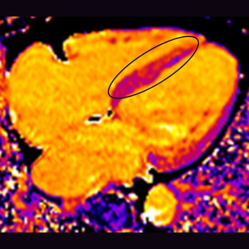
T1 mapping
T1 mapping is one such cardiac MRI technique used at CMIC for the diagnosis and clinical follow up of amyloidosis. T1 mapping measures the relaxation time (T1) of tissues, and maps those times using colour coding. With T1 mapping, subtle changes in myocardial tissue (heart muscle) composition can be detected and quantified, not only identifying the presence of plaques but also quantifying the amyloid burden. This is extremely helpful in monitoring disease progression over time. In the presence of amyloid, T1 relaxation times are typically increased, indicated using ‘hotter’ colours on the colour scale.
Extra-Cellular Volume (ECV) mapping
Extracellular volume (ECV) mapping is another cardiac magnetic resonance imaging (MRI) technique that quantifies the volume fraction of extracellular space within the myocardium (heart muscle). In the context of amyloidosis, ECV mapping provides a quantitative assessment of the space occupied by amyloid plaque within the muscle. Once again, information about the amyloid burden provides valuable insight into the extent and severity of cardiac involvement. ECV mapping can detect early involvement of the myocardium in patients with amyloidosis, even before the onset of overt cardiac symptoms or other imaging features.

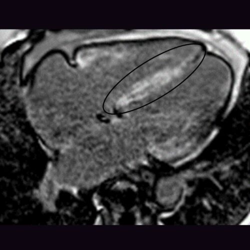
Late Gadolinium enhancement
The injection of a contrast agent called Gadolinium is commonly used in conventional MRI to assess tissue characterisation. Late Gadolinium Enhancement (LGE) imaging is a valuable technique in the diagnosis and monitoring of cardiac amyloidosis as it can demonstrate global diffuse gadolinium enhancement (high signal), an imaging feature of the condition. Not only does LGE allow for the visualisation and localisation of amyloid deposits (useful of disease progression monitoring), but the pattern of LGE in cardiac amyloidosis differs from that seen in other conditions. This distinctive pattern can help differentiate and diagnose cardiac amyloidosis from other causes of myocardial injury.
The future of CMR in the diagnosis and management of cardiac amyloidosis?
Research has made significant progress in recent years, offering insights into the pathophysiology, diagnosis and management of this complex disease. MRI techniques such as T1 mapping, ECV and LGE have been instrumental in detecting myocardial amyloid deposits, quantifying amyloid burden and assessing disease severity. These advanced imaging techniques enable early detection of cardiac involvement, differentiation between amyloid types, and prediction of clinical outcomes.
However, MRI-based research into amyloid is ongoing, and aims to facilitate further development of novel imaging biomarkers for risk stratification, treatment response assessment, and monitoring of disease progression. By leveraging the capabilities of MRI, researchers are advancing general understanding of cardiac amyloidosis and paving the way for improved diagnostic accuracy and therapeutic strategies tailored to individual patients. The Chenies Mews Imaging centre, in partnership with experts in the Amyloidosis field, is proud to be actively involved in Cardiac MRI research.
