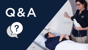

The Chenies Mews Imaging Centre has recently added a new service to its expanding range of specialist MRI services – Fetal MRI. This new and highly specialist magnetic resonance imaging (MRI) service has been established in consultation with City University London, and is led by Dr Kelly Pegoretti Baruteau, a consultant in diagnostic neuroradiology at the National Hospital for Neurology and Neurosurgery in Queen Square. In addition to her extensive experience in using MRI to support the diagnosis of neurological disease in adult patients, Dr Pegoretti Baruteau has a special interest in fetal, neonatal and paediatric neuroimaging.
Q: Firstly Kelly, could you tell us a bit more about your training and background and in particular, what inspired your interest in fetal imaging?
I am Brazilian and studied medicine at the Pontifical Catholic University of Sao Paulo (PUC-SP). I started my Clinical Radiology training in Rio de Janeiro, where I worked for 3 years in various hospitals, including the Sao Vicente de Paulo’s Hospital of Tijuca and the National Institute of Cancer. After that I moved to France to join the training scheme at the Radiology Department of Saint Antoine’s Hospital in Paris.
After 4 years as a radiology trainee I decided to apply for the Neuroradiology Fellowship at the Neuroradiology Department of Toulouse University Hospital. There I gained experience in adults, paediatrics and interventional neuroradiology. This was the first time I experienced working with fetal MRI.
In the UK, I worked as Consultant Neuroradiologist at the Royal London Hospital and as Clinical Research Associate and the Department of Perinatal Imaging and Health, King’s College London, St Thomas’ Hospital. At St Thomas’ Hospital I had the opportunity to work with Professor Mary Rutherford where I gained experience in fetal MRI diagnosis, research and parental counselling.
Currently I work at the National Hospital for Neurorology and Neurosurgery (adult neuroimaging) and at the University College Hospital (fetal and paediatric neuroimaging).
Q: When is fetal MRI typically used?
Antenatal ultrasound is the primary modality for obstetric and fetal imaging. However, MRI is sometimes required as a complementary tool to confirm or exclude abnormal findings suspected on the ultrasound. Sometimes, MRI is also required to give additional information about the developing fetus, most commonly regarding the central nervous system.
Q: What are some of the challenges associated with fetal MRI? Is there anything the mother can do to prepare for their scan?
The biggest challenge to perform a fetal MRI is baby motion. Most of the time this is directly associated with the early gestational ages so in general we will only perform a fetal MRI after 20 weeks of gestation. Mother anxiety may also play a role in the quality of the scan, so we try to ensure she is as comfortable as possible in the scanner. Avoiding caffeine prior to the scan may also help.
Q: Is fetal MRI safe for the mother and baby?
Magnetic Resonance Imaging (MRI) is a non-invasive procedure that does not involve radiation to acquire the images.
Fetal MRI has been performed for more than 30 years and there is no evidence of harm to fetus or mother as long as we ensure that we follow a range of precautionary safety measures before and during the scan.
Q: What should the mother expect during their MRI scan?
After a thorough questionnaire about past medical history and metal check, she will be invited to remove her metallic jewellery (they will be safely kept in a locker) and get changed into a hospital gown, before entering the scanning room. During the procedure she will be laying down on the MRI scanner (with her right side raised with appropriate cushions) and will wear ear plugs to reduce the noises that the MRI scanner will make as it acquire the information we need. This noise is perfectly normal and not hazardous to the fetus. She will also wear headphones to communicate with the radiographer who will be performing the scan. She will handle a buzzer to be triggered anytime she needs to speak with the staff.
Q: What happens after the scan?
After reviewing the images, I will discuss with the parents the first preliminary findings. A formal report will be delivered in the following days as sometimes post-processing of the acquired images may be necessary for a final conclusion.
Q: I am considering having my fetal MRI scan at the Chenies Mews Imaging Centre? Why should I have my scan with you?
As a specialist team, we will strive to deliver the best quality imaging using the best scanners available and also the most humanised service to you and your family. I will use my experience to provide the most accurate diagnosis based on the imaging findings. Following your scan, I will arrange a remote consultation with you so I can discuss the scan results with you, and this will be an opportunity to try to answer to your questions.